INTRODUCTION
Exercise neuroscience is a developing branch of study that includes a wide range of multidisciplinary studies aimed at a better understanding of the brain and body. Internal and external factors such as age, stress, an enriched environment, diet, and physical activity affect the brain and its functions, a phenomenon known as brain plasticity. Brain plasticity refers to the ability of the neuronal system to alter itself structurally, functionally, and molecularly.
In particular, exercise has a substantial physiological, psychological, and social impact on neuroplasticity. What effect does brain activation have and what workout circumstances are employed in the brain it terms of the frequency, intensity, type, and time (FITT) principle? These concerns are crucial for understanding mechanisms that underpin physiological improvements so as to help formulate non-pharmacological treatments to improve brain function.
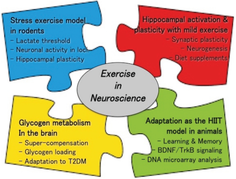
Many studies have been conducted on exercise-induced brain plasticity in animal models, and their findings have been used in studies on health promotion, exercise science, and sports performance. However, the findings cannot be practically applied to humans without first determining their authenticity and mechanism. This study briefly highlights the topics and content benefits of exercise neuroscience.
STRESS EXERCISE MODEL USING RODENTS
This study discusses new advances in the research on neurological benefits of exercise as well as related research [1,2]. Blood lactate and adrenocorticotropic hormone (ACTH) levels rose fast when exercise intensity (running speed) exceeded the lactate threshold (LT), 50-60% of maximum oxygen intake in humans, leading to the production of glucocorticoids(GCs) [3,4]. As a result, activity performed over the LT will be classified as the stress of moderate-intensity stress exercise. However, exercise performed below the LT would be classified as stress-free exercise (mild and/or low-intensity).
Exercise becomes a stressor when it continues to be performed at an intensity over the LT and results in an increase in plasma ACTH, as well as various neurocircuits whose functions are linked to different intensities of running. It is possible that the exercise-induced ACTH response is regulated by arginine vasopressin (AVP) and corticotropin-releasing hormone (CRH). This study also analyzed the activation profile in each brain location at different running speeds based on LT, utilizing c-fos expression as a measure of neuronal activity. The immediate early gene c-fos is believed to have the potential for use as a tool for estimating the activity level of large populations of motor neurons in unrestricted animals [3,5].
The hypothalamus and brain stem are critical regulators of metabolism and stress responses during exercise, as evidenced by these observations. Surprisingly, the hippocampus was stimulated even with low-intensity exercise based on below-LT exercise [6]. The hippocampus is an important part of the humans and other mammalian brains. It is a component of the limbic system that aids in the consolidation of information from short-to long-term memory as well as spatial navigation. Hence, low-intensity exercise may help the hippocampus regenerate neurons and improve cognitive abilities, such as spatial learning and memory.
EXERCISE-INDUCED HIPPOCAMPAL ACTIVATION AND PLASTICITY WITH MOLECULAR MECHANISM
Using a stress exercise model of the brain, we further evaluated the hypothesis of hippocampal activation and neurogenesis. Neurogenesis occurs similarly in the adult hippocampus according to several studies published in the 1990s (thus, it is called adult hippocampal neurogenesis, AHN). The studies showed that exercise boosts the synthesis of brain-derived neurotrophic factor (BDNF)/ tropomyosin receptor kinase B (TrkB) signaling, a protein that helps maintain neuronal cell division, survival, and function, among other things. Their findings suggested that exercise could play a key role in brain activation and the resulting morphological changes [7]. As a result, it has been proposed that modest exercise improves various neural functions via activation of the brain or neuronal activation. This, coupled with the brainŌĆÖs connection to the exercise conditions such as the FITT-VP principle of workout conditions, has attracted considerable attention.
After two weeks of exercise, hippocampal neurogenesis was boosted in the dentate gyrus (DG) under LT. This effect was not observed in the supra-LT group. Even short bursts of below-LT running (moderate exercise) result in a rise in biosynthetic BDNF in the hippocampus, which may help neurons differentiate, survive, and grow [6].
Furthermore, DNA microarray and network analysis indicated a mild exercise (ME)-specific gene inventory encompassing several possible regulators of lipid metabolism, protein synthesis and inflammatory response, and long-term below-LT exercise at 6-weeks, which are recognized as associated with AHN [8]. This evidence may aid in understanding the mechanisms underlying ME-related cognitive gain. However, this effect was not observed in the supra-LT group.
DNA microarray research identified an ME-specific gene inventory encompassing several possible regulators of this positive regulation, and 6-weeks of below-LT exercise showed the ability to improve AHN. This information could help researchers determine how ME causes cognitive gain. Astaxanthin (ASX) supplementation in exercise nutrition has been added to this concept. A particular molecular substrate may improve spatial memory when ASX is paired with modest exercise-enhanced AHN [9]. Based on these findings, we decided to investigate the optimal exercise intensity for hippocampal plasticity.
Additionally, the muscle secretory factor cathepsin B (CTSB) is important for the cognitive and neurogenic benefits of running. Recombinant CTSB application enhanced the expression of BDNF and doublecortin (DCX) in adult hippocampal progenitor cells [10]. Exercise increased CTSB levels in the mouse gastrocnemius muscle and plasma. Proteomic analysis revealed elevated levels of CTSB in conditioned medium derived from skeletal muscle cell cultures treated with the AMP-kinase agonist 5-aminoimidazole-4-carbozamide-1-╬▓-D-ribonucleoside(AICAR) [11]. Therefore, physical activity can be an encouraging intervention for brain restoration through neuronal plasticity [12].
GLYCOGEN METABOLISM IN THE BRAIN
The brain is a high-energy organ that uses approximately 20% of the bodyŌĆÖs calories and requires regular food intake to function properly. The brain uses lactate as an energy source for neuromodulation. Lactate is obtained by the brain in two ways: (a) externally via the bloodstream, and (b) internally via astrocytes via glycolysis and/or glycogenolysis. Astrocyte-stored glycogen is a key source of lactate generation in the brain [13].
Lactate is created by astrocyte-stored brain glycogen and transferred by monocarboxylate transporters (MCTs) to maintain neuronal activities such as hippocampus-regulated memory formation. Although activity activates brain neurons, the role of astrocytic glycogen in the brain during exercise remains unclear. This study examined the energetic role of astrocytic glycogen in the brain during exercise to sustain endurance capacity via lactate transfer, as muscle glycogen fuels active muscles during exercise.
Through monocarboxylate transporters, astrocyte glycogen plays an energy role in maintaining neuronal function in the brain after prolonged exercise [13]. Lactate, which is generated from astrocyte glycogen, provides energy to the brain, allowing it to retain its endurance during hard exercise. Glycogen-maintained brain ATP levels could be a protective mechanism for neurons in a state of exhaustion [14]. Therefore, we investigated whether glycogen loading (GL) increases glycogen levels in the brain or muscle using a rat model. According to our findings, GL increases glycogen levels in the hippocampus, hypothalamus, and muscle [15].
In addition, rats with type 2 diabetes mellitus (T2DM) displayed abnormalities in astrocyte-neuron lactate shuttle (ANLS)-related glucose metabolism in the hippocampus, higher glycogen levels and lower MCT2 protein levels. Memory impairment can be improved with chronic exercise in the form of an ANLS adaptation [16,17]. In both the hippocampus and hypothalamus, T2DM rats exhibit reduced MCT2 protein levels and greater glycogen levels, which are related to HbA1C levels, indicating a probable dysregulation of glucose metabolism at both brain loci in T2DM [18].
As a result, similar to skeletal muscle, glycogen supercompensation in the brain occurs after intense activity. Furthermore, the role of the ANLS in the etiology of brain dysfunction in patients with obesity and T2DM should be studied. Furthermore, brain glycogen levels, particularly in the hippocampus, have increased, implying a physiological strategy to boost hippocampal function.
ADAPTATION AS THE HIGH-INTENSITY TRAINING MODEL IN ANIMALS
According to the American College of Sports Medicine, high-intensity interval training (HIIT) is a consistently high-ranking prediction trend. Short bursts of high-intensity, high-effort training (usually 20 to 90 s) are followed by a short rest time or low-intensity recovery in HIIT workouts [19]. HIIT has the same effect as endurance exercise on aerobic capacity, mitochondrial fatty acid oxidation, and cardiovascular risk [20,21]. As an animal model corresponding to HIIT, voluntary resistance wheel running (VRWR), noted on a voluntary running wheel for a given load, is a suitable workout model for increasing work levels as energy expenditure without using physical or psychological stressors such as an electrical shock, weight vest, food deprivation, and duration [22,23].
To address this, we set out to create an animal model of strength training, examine whether HIIT affects cognitive functioning, and unravels the underlying mechanisms. Our analysis showed that voluntary resistance wheel running could improve brain function (VRWR) [24,25]. Although acute and chronic VRWR replicate the effects of traditional wheel-running on learning and memory by increasing high-energy expenditure with the load but not increasing running distance (half of the control), the downstream transcription factor CREB, induces various gene transcriptions related to cell survival and neuroplasticity [24].
In addition, both acute and chronic VRWR caused hippocampal changes in rats, including neurogenesis [25, 26]. They also produced a list of newly altered gene expressions and observed modifications in some transcriptional pathways in the hippocampi of VRWR rats [27]. Thus, HIIT is comparable to VRWR and intermittent exercise in terms of effectiveness. It benefits brain plasticity and produces shorter distances but higher effort levels as a high-efficiency exercise.
CONCLUSION AND PERSPECTIVES
In animal studies, exercise has been shown to have a favorable effect on the brain. Physical activity is beneficial for brain health. Exercise stimulates molecules, causes structural changes, and aids behavioral development. By highlighting various features of the neural system, physical activity promotes neuroplasticity in both healthy and pathological conditions. Although there has been a large accumulation of evidence on exercise nesuroscience, further studies will require thorough verification and clinical intervention trials involving humans. The truth and mechanism must be established before it can be applied to humans. Exercise-induced molecular, functional, and structural alterations in rodents and humans may affect performance. Overall, the goal of exercise neuroscience research is to learn more about the physiological and psychological effects of exercise on human health and performance.


